
The affected vessel may also enlarge 9.Īcute pulmonary thromboemboli can rarely be detected on non-contrast chest CT as intraluminal hyperdensities 12.ĭual-energy CT holds much promise for the diagnosis and prognosis of PE (see cases 33 and 34). Typically the embolus makes an acute angle with the vessel, in contrast to chronic emboli. When the artery is viewed in its axial plane the central filling defect from the thrombus is surrounded by a thin rim of contrast, which has been called the Polo Mint sign.Įmboli may be occlusive or non-occlusive, the latter is seen with a thin stream of contrast adjacent to the embolus. Sensitivity and specificity of chest x-ray signs 1:ĬT pulmonary angiography (CTPA) will show filling defects within the pulmonary vasculature with acute pulmonary emboli.

It is used to assess for differential diagnostic possibilities such as pneumonia and pneumothorax rather than for the direct diagnosis of PE.ĭescribed chest radiographic signs include: Plain radiographĬhest radiography is neither sensitive nor specific for a pulmonary embolism. Overall, there is a predilection for the lower lobes. Classificationĭepends to some extent on whether it is acute or chronic. Patients are treated with anticoagulants while awaiting the outcome of diagnostic tests 4. In patients with a high probability clinical assessment, a D-dimer test is not helpful because a negative D-dimer result does not exclude pulmonary embolism in more than 15%. raised D-dimer is seen with PE but has many other causes and is, therefore, non-specific: it indicates the need for further testing if pulmonary embolism is suspected 4.normal D-dimer has almost 100% negative predictive value (virtually excludes PE): no further testing is required.non-HIV matched controlsĭ-dimer (ELISA) is commonly used as a screening test in patients with a low and moderate probability clinical assessment, on these patients: malignancy: including multiple myeloma 23.S IQ IIIT III pattern: this refers to a deep S wave in lead I, Q wave and T wave inversion in lead III this sign is seen as classical of PE but is, in fact, poor in terms of both sensitivity and specificity as a sign of PE.

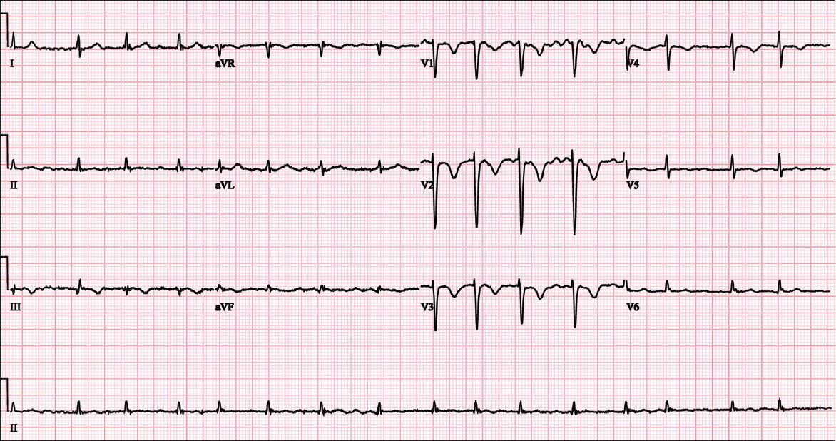
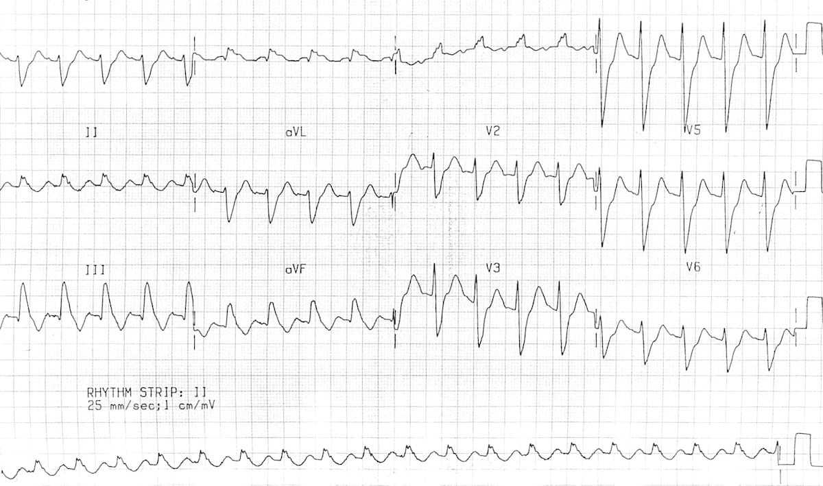
simultaneous T-wave inversion in lead III and V 1 has been repeatedly shown to have strong sensitivity (90%), specificity (97%), positive predictive value (92%), negative predictive value (96%) and overall predictive accuracy of 95% 25,26.T-wave inversion in the right precordial leads +/- the inferior leads is seen in up to 34% of patients and is associated with high pulmonary artery pressures 25.

incomplete or complete right bundle branch block.sinus tachycardia: the most common abnormality.tenderness to palpation along the deep venous systemĬlinical decision rules, in conjunction with physician gestalt and estimated pretest probability of disease, may serve as a supplement in risk stratification:.prominent superficial collateral vessels.asymmetric pitting lower extremity edema.clinical signs of deep venous thrombosis (DVT).The physical exam may reveal suggestive features such as: The patient may report a history of recent immobilization or surgery, active malignancy, hormone usage, or a previous episode of thromboembolism. the presence or absence of hemodynamic compromise.Classification of a pulmonary embolism may be based upon:


 0 kommentar(er)
